Faculty Profiles
- Faculty Leader Profiles
- Recruiting Faculty
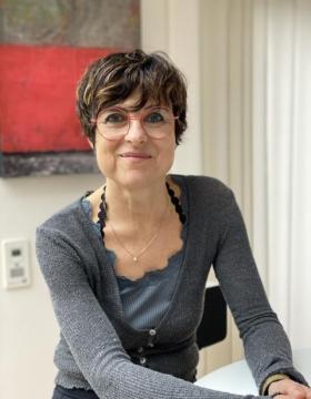
Prof. Dr. Amparo Acker-Palmer
Goethe University Frankfurt - Institute of Cell Biology and NeuroscienceCell Biology
Research Mission
Over a decade my group has been working on the structural and functional parallelism between nervous and vascular development and plasticity. We direct our efforts to combine the research of basic molecular mechanisms during physiological processes with the investigation of pathological conditions in the brain. Our studies elucidate the molecular signaling cascades important for cellular behavior and intercellular communication in the brain.
Experimental Techniques
Our diverse methodological repertoire includes biochemical and cell biological methods, in vitro, ex vivo (e.g. retina explants), and in vivo mouse and zebrafish technologies, gene editing with CRISPR, scRNAseq, electrophysiology, and state of the art imaging techniques like expansion microscopy, confocal and multi-photon microscopy.

Dr. Martin Beck
MPI of Biophysics - Molecular SociologyStructural Biology
Research Mission
Functional cellular modules have often been characterised in vitro. However, relatively little is known about their interplay and spatio-temporal arrangement in the context of living cells, i.e. their 'molecular sociology'. We use integrative, in situ structural biology techniques to study the structure, function and assembly of very large macromolecular complexes in their native environment.
Experimental Techniques
We rely on a diverse methodological repertoire, including cryo-EM, structural proteomics, biochemistry, imaging, animal model systems and computational modeling

Dr. Ramachandra M Bhaskara
Goethe University Frankfurt - Institute of Biochemistry II (IBC2)Biophysics
Research Mission
Our team focuses on tackling challenging problems at the interface of Cell Biology, Physics, and, Data Sciences, especially to decipher cellular patterns and processes at the mesoscale. We develop and apply theoretical modeling, novel computer simulations, and, data integration methods to understand complex biological systems.
Experimental Techniques
We employ Integrative Modeling and AI methods to decode relationships between protein sequence, structure, function & evolution. We study protein-induced membrane mechanics to reveal molecular mechanisms at cellular and organellar membranes (e.g. Cellular homeostatic pathways) by capturing the dynamics of biomolecular systems at various temporal and spatial scales. We employ data science approaches to integrate disparate datasets effectively.
Selected Publications
Intrinsically disordered region amplifies membrane remodeling to augment selective ER-phagy.
Proc. Natl. Acad. Sci. USA. 2024
An atypical GABARAP binding module drives the pro-autophagic potential of the AML-associated NPM1c variant.
Cell Reps. 2023
Curvature induction and membrane remodelling by FAM134B reticulon homology domain.
Nat. Commun. 2019

Dr. Roberto Covino
Frankfurt Institute for Advanced Studies (FIAS)Biophysics
Research Mission
In cellular membranes, lipids and membrane proteins self-assemble into heterogeneous and dynamic complexes. Membranes tell proteins how to move and arrange, and proteins reshape membranes in return. The dynamic modulation of this choreography is at the origin of many regulatory programs in the cell. Our goal is to quantitatively understand complex molecular mechanisms in cellular membranes in terms of general physical principles.
Experimental Techniques
We use state-of-the-art molecular dynamics simulations and develop innovative computational methods integrating AI with physics-based modeling. Our approach is highly interdisciplinary, and we work in a constant exchange with experiments. Our primary focus is to decipher the molecular activation mechanism of the unfolded protein response, a fundamental homeostatic pathway central in health and disease.
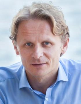
Prof. Dr. Ivan Dikic
Goethe University Frankfurt - Institute of Biochemistry II (IBC2)Biochemistry
Research Mission
Cellular homeostasis is achieved, among others, by tight regulation of the two major degradation pathways – the ubiquitin-proteasome system (UPS) and autophagy. It is critical to understand the structure and function of individual components within each of these pathways, as any disruption to either of the processes can lead to neurodegenerative diseases, cancer and other pathologies. To study functional and structural properties of components of
Experimental Techniques
To tackle our research questions, we rely on a wide variety of biological and chemical techniques, including biochemistry, proteomics, cell imaging, X-ray crystallography, Cryo-EM and animal model systems.

Prof. Dr. Dorothee Dormann
Johannes Gutenberg University Mainz - Institute of Molecular Biology (IMB)Neurodegeneration
Research Mission
Our research focuses on the molecular mechanisms of RNA-binding protein (RBP) dysfunction in neurodegenerative diseases, most notably the currently incurable disorders ALS (amyotrophic lateral sclerosis), FTD (frontotemporal dementia), and Alzheimer's disease. We aim to understand how specific, disease-linked RBPs, such as TDP-43 and FUS, mislocalize and aggregate in these disorders, to provide a mechanistic basis for new therapeutic strategies.
Experimental Techniques
We dissect the complex pathomechanism using a reductionist approach based on in vitro reconstitution and cellular experiments. We use recombinant proteins, protein/RNA biochemistry and biophysics to understand molecular events. As cellular models, we mostly use mammalian cell lines. We use techniques such as confocal microscopy, live cell imaging, protein-RNA interaction and phase separation assays, and employ genomics and proteomics approaches.
Selected Publications
Disease-linked TDP-43 hyperphosphorylation suppresses TDP-43 condensation and aggregation
EMBO Journal 2022
Phase Separation of FUS Is Suppressed by Its Nuclear Import Receptor and Arginine Methylation
Cell 2018
Adding intrinsically disordered proteins to biological ageing clocks
Nature Cell Biology 2024

Prof. Dr. Volker Dötsch
Goethe University Frankfurt - Institute of Biophysical ChemistryBiophysics
Research Mission
Our goal is to contribute to the understanding of biological structure, function and interaction at a molecular level. Among our main concerns are the characterization of the transcription factor p63, homolog of the famous tumor suppressor protein p53, the investigation of membrane proteins and the study of the molecular basis for autophagy.
Experimental Techniques
We utilize a wide range of biological, chemical and physical state-of-the-art techniques such as NMR, cell culture, cell-free protein production, genetic engineering, EPR, chemical labelling, and many more.
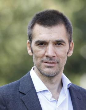
Prof. Dr. Achilleas Frangakis
Goethe University Frankfurt - Institute of BiophysicsBioimaging
Research Mission
Cell adhesion is an essential process that allows for cells to attach and interact with other cells and their environment in order to perform a multitude of tasks. Although adhesion is directly related to infection processes and disease, it is currently only poorly understood. Our mission is to study and decipher the adhesion mechanisms of both eukaryotic and bacterial cells.
Experimental Techniques
We use a large arsenal of different technologies to complement our primary methodologies of cryo-electron tomography, image processing and machine learning. Our main model system to study bacterial adhesion is M. pneumoniae & genitalium. For eukaryotic systems we utilise various endothelial and epithelial cell lines.
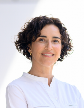
Prof. Dr. Ana García Sáez
MPI of BiophysicsBioimaging
Research Mission
Our department aims at understanding the underlying physical principles and molecular mechanisms that govern membrane organization and dynamics. A key open question is how membrane behavior and signaling are coordinated by the interplay between molecular components and the membrane properties. We focus our research on the mitochondrial alterations during apoptosis as well as on membrane permeabilization as a common theme in regulated cell death.
Experimental Techniques
We have established a multi-disciplinary department that develops new microscopy tools and combines biophysics, biochemistry and cell biology in reconstituted minimal systems and in single cells to analyze mitochondrial permeabilization and additional alterations during apoptosis from a quantitative perspective. We also apply these methods to study the molecular mechanisms of membrane permeabilization that execute other forms of cell death.
Selected Publications
The interplay between BAX and BAK tunes apoptotic pore growth to control mitochondrial DNA-mediated inflammation
Molecular Cell 2022
DRP1 interacts directly with BAX to induce its activation and apoptosis
EMBO Journal 2022
BCL-2-family protein tBID can act as a BAX-like effector of apoptosis
EMBO Journal 2022

Prof. Dr. Clemens Glaubitz
Goethe University Frankfurt - Institute of Biophysical ChemistryBiophysics
Research Mission
The Glaubitz Lab resolves the molecular mechanisms of membrane proteins. In addition, we study the effect of lipid-protein interactions and factors influencing membrane order, dynamics and polymorphism. We are especially intersted in mechanisms by which membrane proteins maintain complex membrane systems such as those found in Gram-negatve bacteria.
Experimental Techniques
Our methodological approach is centered around solid-state NMR (high field, ultra fast and dynamic nucelar polarisation MAS-NMR) complemented by a range of biochemical approaches. Current research covers novel photoreceptors, transport proteins, lipid regulators and GPCRs. We also explore the use of light to manipulate and control native and artificial processes within membranes.
Selected Publications
How Photoswitchable Lipids Affect the Order and Dynamics of Lipid Bilayers and Embedded Proteins.
J Am Chem Soc 2021
Probing the photointermediates of light-driven sodium ion pump KR2 by DNP- enhanced solid-state NMR
Science Advances 2021
Photocycle-dependent conformational changes in the proteorhodopsin cross-protomer Asp-His-Trp triad revealed by DNP-enhanced MAS-NMR
Proc Natl Acad Sci USA 2019
Unexplored nucleotide binding modes for the ABC exporter MsbA
J Am Chem Soc 2018
Coupled ATPase-adenylate kinase activity in ABC transporters
Nature Communications 2016

Prof. Dr. Alexander Gottschalk
Goethe University Frankfurt - Institute of Biophysical ChemistryNeurobiology
Research Mission
We want to understand how neuronal circuits orchestrate behavior, and how signal transmission occurs within these circuits, made from chemical, electrical and neuromodulatory synapses. We study this in Caenorhabditis elegans, with 302, mostly "hard-wired" neurons, and the zebrafish, as a complex vertebrate model. We are interested in the subcellular organization of synapses and of molecular machines enabling them to orchestrate neurotransmission.
Experimental Techniques
We use genetics / molecular biology to study and manipulate genes. By electrophysiology, calcium- and voltage-imaging, we study cellular activity in circuits and at the neuromuscular junction, and we use electron microscopy to analyze synaptic ultrastructure. All these are combined with optogenetics, enabling to analyze and quantify resulting behavior. Last, we develop specific optogenetic tools. Models: Caenorhabditis elegans Danio rerio

Dr. Bernhard Hampölz
MPI of Biophysics - Molecular SociologyCell Biology
Research Mission
Our aim is to understand how macromolecular complexes and in particular the Nuclear Pore Complex (NPC) assemble in the context of animal development. To that end we use embryos and the female germline of the fruitfly Drosophila melanogaster to delineate principles of how NPCs form in a multicellular organism and how their assembly relies on the major regulatory inputs of early development.
Experimental Techniques
Our methodological repertoire is broad and spans techniques as diverse as cultivating and crossing fruitfly lines (aka “Fly pushing”) and in vitro biochemistry. This spectrum contains Drosophila genetics, general cell biological techniques, organ dissection, plastic and Cryo-EM (including CLEM). Our core technology is imaging of living Drosophila embryos, ovaries and larval brains.
Selected Publications
Nuclear Pores Assemble from Nucloporin Condensates During Oogenesis
Cell 2019
Pre-assembled Nuclear Pores Insert Into the Nuclear Envelope During Early Development
Cell 2016
The small GTPase Ran defines Nuclear Pore Complex Asymmetry
Cell 2025
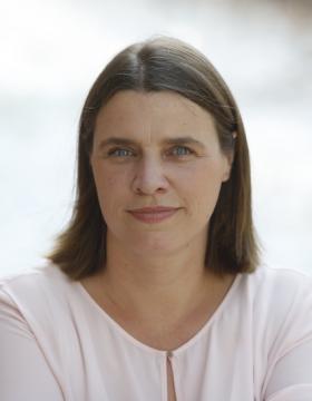
Prof. Dr. Inga Hänelt
Goethe University Frankfurt - Institute of BiochemistryBiochemistry
Research Mission
Proteins involved in substrate translocation across biological membranes are diverse and the variety that nature has evolved to control substrate flux absolutely fascinates us. A major focus of my group is on bacterial membrane transport systems and, in particular, potassium transport systems that are essential for the survival of a single cell and a bacterial community, namely biofilms.
Experimental Techniques
Our lab applies a variety of methods, including biochemistry, structural biology, EPR spectroscopy, microbiology and electrophysiology for understanding the molecular principles of membrane transport proteins.
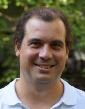
Prof. Dr. Mike Heilemann
Goethe University Frankfurt - Institute of Physical and Theoretical ChemistryBioimaging
Research Mission
Our mission is to develop super-resolution fluorescence microscopy into an "optical omics" technology for structural cell biology. We develop imaging tools for quantitative microscopy with single-protein resolution, along with novel computational tools for microscopy. We investigate how proteins organize in cells and tissue, and how this organization correlates with protein function.
Experimental Techniques
super-resolution microscopy (dSTORM, PALM, DNA-PAINT, STED, SOFI), live cell microscopy, deep-learning assisted microscopy, chemical biology, fluorescence spectroscopy
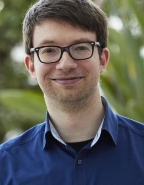
Prof. Dr. Eric Helfrich
Goethe University Frankfurt - Institute for Molecular Bio ScienceSynthetic & Chemical Biology
Research Mission
Our research focuses on unraveling the blueprints involved in the biosynthesis of complex bacterial natural products. We develop bioinformatic tools to explore the hidden biosynthetic diversity within bacterial genomes and investigate the biosynthesis of these intricate metabolites. Our work delves into the role of compartmentalisation within the biosynthetic process and aims to characterise biosynthetic enzymes and unprecedented transformations.
Experimental Techniques
Our interdisciplinary team works at the interface of bioinformatics, molecular and synthetic biology, and analytical chemistry. We develop machine learning-based tools to generate hypotheses, which we validate in the lab using the latest techniques in the fields of molecular and synthetic biology to produce complex natural products. Moreover, we use HPLC, mass spectrometry and NMR to characterise the bacterial natural products generated.
Selected Publications
Atropopeptides are a Novel Family of Ribosomally Synthesized and Posttranslationally Modified Peptides with a Complex Molecular Shape
Angewandte Chemie International Edition 2022
Machine learning-based exploration, expansion and definition of the atropopeptide family of ribosomally synthesized and posttranslationally modified peptides
bioRxiv 2023
Evolution of combinatorial diversity in trans-acyltransferase polyketide synthase assembly lines across bacteria
Nature Communications 2021
Specialized Metabolites Reveal Evolutionary History and Geographic Dispersion of a Multilateral Symbiosis
ACS Central Science 2021

Prof. Dr. Gerhard Hummer
MPI of Biophysics - Theoretical BiophysicsBiophysics
Research Mission
We use molecular dynamics simulations and integrative modeling to study biological systems from molecular to organellar scales. Focus areas include the dynamics of the nuclear pore complex; membrane remodeling in cell homeostasis, autophagy and viral infection; and membrane channel and transporter function. To tackle issues of size and complexity in molecular dynamics simulations at organellar scales, we develop novel simulation and AI methods.
Experimental Techniques
We use a wide range of computational and theoretical techniques including molecular dynamics simulations; integrative modeling; artificial intelligence / machine learning; statistical physics; computational physics; theoretical chemistry; quantum chemistry; computational chemistry; bioinformatics; high-performance computation; kinetic modeling; stochastic processes; and Bayesian statistics.
Selected Publications
Artificial intelligence reveals nuclear pore complexity
bioRxiv 2022
Global structure of the intrinsically disordered protein tau emerges from its local structure
JACS Au 2022
Membrane fusion and drug delivery with carbon nanotube porins
Proc. Natl. Acad. Sci. U.S.A. 2021
FAM134B-RHD protein clustering drives spontaneous budding of asymmetric membranes
J. Phys. Chem. Lett. 2022
In situ structural analysis of SARS-CoV-2 spike reveals flexibility mediated by three hinges
Science 2021
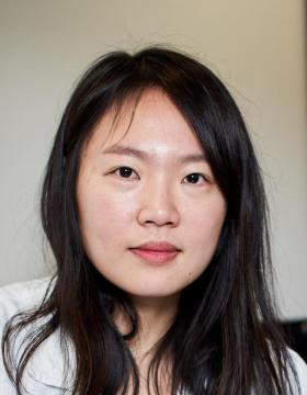
Dr. Eugene Kim
MPI of BiophysicsBiophysics
Research Mission
Cellular DNA is highly organized not only to fit within the cells, but also to regulate genomic processes. Yet, what determines DNA organization and how it impacts on cellular function remain largely unknown. We study biochemical and physical processes underlying chromosome organization by monitoring their dynamics at the single-molecule level with high spatiotemporal resolution.
Experimental Techniques
We employ single-molecule fluorescence imaging, single-molecule force spectroscopy, and correlative light and electron microscopy techniques, in combination with analytical biochemistry tools.

Prof. Dr. Stefan Knapp
Goethe University Frankfurt - Institute of Pharmaceutical ChemistryStructural Biology
Research Mission
We are probing cellular signalling pathways by designing highly selective chemical tools that allow us to modulate endogenous cellular proteins, to visualize them and to study molecular assemblies.We would like to understand how cellular signalling pathways are perturbed in diseases and how we can reverse deregulated signalling events for the development of new therapies.
Experimental Techniques
We predominantly use (high throughput) crystallography for rationally designing chemical tools but have also started to use single particle cryo-EM.
Selected Publications
Structure-Based Design of Selective Salt-Inducible Kinase Inhibitors.
J Med Chem. 2021
Structure-kinetic relationship reveals the mechanism of selectivity of FAK inhibitors over PYK2
Cell Chem. Bio. 2021
PROTAC-mediated degradation reveals a non-catalytic function of AURORA-A kinase
Nature Chem. Biol. 2021
Trends in kinase drug discovery: targets, indications and inhibitor design
Nat Rev Drug Discov 2021
Structure of LRRK2 in Parkinson's disease and model for microtubule interaction
Nature 2020
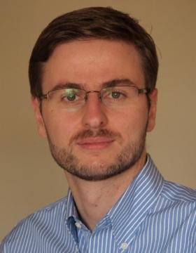
Dr. Jürgen Köfinger
MPI of Biophysics - Theoretical BiophysicsBiophysics
Research Mission
Mechanistic insight into biomolecular processes relies on resolving structures, dynamics, and interactions of biomolecules and their complexes. In many cases, a single experimental or theoretical method is not capable to complete this task. Thus, we develop and apply theoretical tools to integrate information from different sources, i.e., from experiments, bioinformatics, biomolecular modeling, and molecular simulations.
Experimental Techniques
We use statistical mechanics, statistical data analysis, and information theory to integrate experimental data and biomolecular simulations. In ensemble refinement we use experimental data with low resolution or low structural information content (SAXS, WAXS, DEER, ...) to refine thermodynamic structural ensembles of dynamic biomolecules and biomolecular complexes. We usually, but not always, generate these ensembles in molecular simulations.
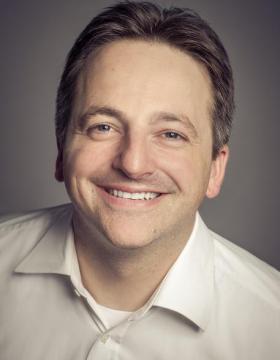
Dr. Julian Langer
MPI of BiophysicsProteomics
Research Mission
My research focuses on the development of mass spectrometry-based methods for studying individual membrane proteins, purified sub-proteomes and complex prokaryotic and eukaryotic systems. We make use of the unique data that mass spectrometry can provide for structural biology and in functional studies, and customize our methods to analyze challenging targets that are often inaccessible with conventional approaches.
Experimental Techniques
Our techniques include direct, digest-free sequencing of membrane proteins, HDX-MS, functional assays on membrane protein complexes, non-canonical amino acid tagging, shotgun and phospho-proteomics as well as proteome turnover studies and MALDI-Imaging. We work on a wide range of target proteins and proteomes, ranging from identifying small membrane proteins in prokaryotes to studying neuronal cell type-specific proteome dynamics in vivo.
Selected Publications
The proteomic landscape of synaptic diversity across brain regions and cell types
Cell 2023
Detection of Known and Novel Small Proteins in Pseudomonas stutzeri Using a Combination of Bottom-Up and Digest-Free Proteomics and Proteogenomics
Analytical Chemistry 2023
An abundance of free regulatory (19 S) proteasome particles regulates neuronal synapses
Science 2023
Essential protein P116 extracts cholesterol and other indispensable lipids for Mycoplasmas
Nature Structural and Molecular Biology 2023
A Practical Guide to Small Protein Discovery and Characterization Using Mass Spectrometry
Journal of Bacteriology 2021
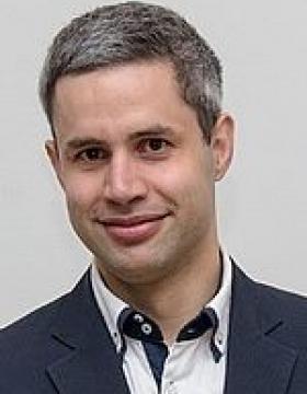
Prof. Dr. Edward A Lemke
Johannes Gutenberg University Mainz - Institute of Molecular Biology (IMB)Biophysics
Research Mission
We focus on studying intrinsically disordered proteins (IDPs), which constitute up to 50% of the eukaryotic proteome. IDPs are found in many vital biological processes, such as nucleocytoplasmic transport, transcription and gene regulation. The ability of IDPs to exist in multiple conformations is considered a major driving force behind their enrichment during evolution in eukaryotes.
Experimental Techniques
Using a question-driven, multidisciplinary approach paired with novel tool development in miroscope engeenering, chemical biology, synthetic biology and microfludics, we have made major strides in understanding the biological dynamics of such systems from the single molecule to the super resolved whole cell level.
Selected Publications
Visualizing the disordered nuclear transport machinery in situ
Nature 2023
Dual film-like organelles enable spatial separation of orthogonal eukaryotic translation.
CELL 2021
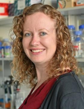
Dr. Melanie McDowell
MPI of BiophysicsStructural Biology
Research Mission
Almost all eukaryotic membrane proteins start their life in the cytosol and must journey to the cellular membrane where they function. Multiple pathways are employed to recognise different classes of membrane proteins, deliver them to the correct membrane and insert them into the lipid bilayer. We strive to understand the protein factors involved in these pathways at the endoplasmic reticulum, the primary destination for most membrane proteins.
Experimental Techniques
We use an integrative approach to study protein structure, interactions and function. Techniques include cryo-EM, x-ray crystallography, biophysical methods, in vitro biochemical reconstitution and pull-outs of native protein complexes from model organisms such as yeast and Chaetomium thermophilum.
Selected Publications
Structural and molecular mechanisms for membrane protein biogenesis by the Oxa1 superfamily
Nature Structural and Molecular Biology 2021
Structural basis of tail-anchored membrane protein biogenesis by the GET insertase complex
Molecular Cell 2020
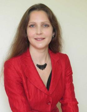
Prof. Dr. Nina Morgner
Goethe University Frankfurt - Institute of Physical and Theoretical ChemistryPhysical Chemistry
Research Mission
Cellular function is generally controlled by non-covalent interactions between proteins and other biomolecules. We investigate such interactions, molecular machines and their assembly by means of native mass spectrometry (MS). Where current technology is not sufficient to answer we further develop mass spectrometric instrumentation.
Experimental Techniques
Our research makes use of two different non-covalent MS methods. One is the commercially available nESI-MS method, the other LILBID MS, which we develop in house.
Selected Publications
Bacterial F-type ATP synthases follow a well-choreographed assembly pathway.
Nature communications 2022
LILBID laser dissociation curves: a mass spectrometry-based method for the quantitative assessment of dsDNA binding affinities.
Scientific reports 2020
Structural rearrangement of amyloid-β upon inhibitor binding suppresses formation of Alzheimer’s disease related oligomers
eLife 2020
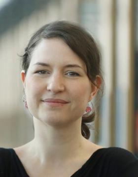
Prof. Dr. Michaela Müller-McNicoll
Goethe University Frankfurt - Institute for Molecular Bio ScienceGene Regulation
Research Mission
RNA binding proteins with low complexity domains (LCDs) often assemble nuclear compartments or large RNA-protein particles under stress. We study how RNA-protein interactions organize subcellular architecture and coordinate the regulation of gene expression during cellular stress and differentiation. Our goal is to understand how different steps in gene expression are interconnected in time and separated in space.
Experimental Techniques
Using pluripotent cells as a model system, we combine various methods including smRNA FISH, imaging, protein/RNA Biochemistry, iCLIP, Ribo-seq, metabolic labelling, reporter gene studies, mass spectrometry, CRISPR mutagenesis and RNA bioinformatics to obtain mechanistic insights. We also develop new methods and assays to get a quantitative and high-resolution picture of gene expression in the context of a compartmentalized cell.
Selected Publications
Poison exon splicing of SRSF6 regulates nuclear speckle dispersal and the response to hypoxia.
Nucleic Acids Research 2023
SRSF3 and SRSF7 affect 3’UTR length in opposite directions through suppression or activation of proximal poly(A)-sites and regulation of CFIm levels.
Genome Biology 2021
SRSF7 maintains its homeostasis through the expression of Split-ORFs and nuclear body assembly
Nature Structural Molecular Biology 2020
Cellular differentiation state modulates the export activity of SR proteins.
Journal of Cell Biology 2017
hGRAD: A versatile "one-fits-all" system to acutely deplete RNA binding proteins from condensates
Journal of Cell Biology 2024
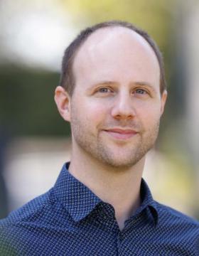
Prof. Dr. Christian Münch
Goethe University Frankfurt - Institute of Biochemistry II (IBC2)Cell Biology
Research Mission
Cells respond to stress with highly coordinated and dynamic responses. Their full complexity and spatio-temporal control remains poorly understood. We focus on a range of stresses - (mitochondrial) protein misfolding, infection, cancer - to study their effects on a systems biology/medicine level. We aim to define the molecular details of stress responses, their roles in diseases, and potential avenues for therapeutic intervention.
Experimental Techniques
We employ biochemical, cell biological, genetic, and proteomics approaches to comprehensively monitor the multiple facets of stress responses. To grasp dynamic, cell-wide changes, we develop proteomics methods, such as for translation & degradation. We then apply computational approaches on our multi-level data to gain integrated systems information. Our work largely focuses on mammalian cell culture systems and clinical models.
Selected Publications
A cytosolic surveillance mechanism activates the mitochondrial UPR
Nature 2023
Global mitochondrial protein import proteomics reveal distinct regulation by translation and translocation machinery
Molecular Cell 2022
Protein import motor complex reacts to mitochondrial misfolding by reducing protein import and activating mitophagy
Nature Commun 2022
Proteomics of SARS-CoV-2-infected host cells reveals therapy targets
Nature 2020
Functional Translatome Proteomics Reveal Converging and Dose-Dependent Regulation by mTORC1 and eIF2α
Molecular Cell 2020

Dr. Bonnie Murphy
MPI of BiophysicsStructural Biology
Research Mission
Our group is focused on gaining a better understanding of the structure and especially the mechanism of redox and metalloproteins across the tree of life. We also work to develop methods for elemental mapping in macromolecular complexes, combining analytical techniques typically used for dose-tolerant inorganic samples with the image processing tools of single-particle analysis.
Experimental Techniques
We use single-particle cryo-EM as a primary tool in the lab, complemented by biochemical and electrochemical tools. We use additional analytical capabilities of the microscope in STEM mode (eg. 4D-STEM, EELS analysis), giving us richer information about our sample.
Selected Publications
see our website / Google scholar page for up-to-date publication list
2026
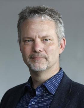
Prof. Dr. Klaas Martinus Pos
Goethe University Frankfurt - Institute of BiochemistryBiochemistry
Research Mission
We look into the molecular detail of antibiotic efflux pump and lipid transport machineries and how these pumps can provide resistance against many antibiotics. We also research on efflux pump inhibitors, which can be used in combination with existing antibiotics to recover their effectiveness.
Experimental Techniques
We use X-ray crystallography and single-particle Cryo-EM to obtain molecular insight into the mechanisms of antibiotic binding and transport. With biophysical and biochemical methods, we quantify the effectiveness of the efflux pump inhibitors and gain insight into antibiotic interaction with the efflux pump
Selected Publications
Pyridylpiperazine efflux pump inhibitor boosts in vivo antibiotic efficacy against K. pneumoniae.
EMBO Mol Med 2024
Pyridylpiperazine-based allosteric inhibitors of RND-type multidrug efflux pumps
Nat Commun 2022
Allosteric drug transport mechanism of multidrug transporter AcrB
Nat Commun 2021

PhD. Uris Lianne Ros Quincoces
MPI of BiophysicsCell Biology
Research Mission
Cell death is commonly a consequence of the sustained disturbance in membrane integrity. Our mission is to understand the connection between cell death, membrane damage and cell signaling. Our vision is built on the hypothesis that cells integrate the response to different forms of membrane damage into common repair and response principles and that the interconnection of these processes determines how cells and tissues cope with danger signals.
Experimental Techniques
We use state-of-the art membrane biophysics, structural and cell biology, biochemistry, and quantitative light microscopy. The combination of these techniques allows us to obtain mechanistic information about the integration of cell signaling and different forms of membrane damage. We exploit this knowledge for the development of new therapies against cell-death related diseases.
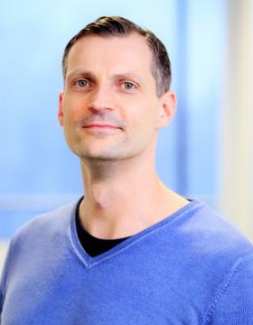
Dr. Andreas Schlundt
Goethe University Frankfurt - Institute for Molecular Bio ScienceStructural Biology
Research Mission
We study RNA-protein complexes that decide about mRNA turnover and thus form a major regulatory parameter in cell homeostasis. Complex formation is not only driven by protein abundance, but also by the availability, structure and stability of the targeted RNA elements to be found in mRNA untranslated regions. We seek to understand, on an atom-resolved level, the structure and mechanisms of mRNA-protein networks that encode the mRNA's fate.
Experimental Techniques
We are using integrated structural biology which combines a broad set of methods from biochemistry, biophysics and structural biology. Our foucs is set to solution methods, in particular NMR and SAXS. My lab has an expertise in the recombinant production of proteins, primarily in bacteria, and the in-vitro transcription of RNAs.
Selected Publications
Structural basis for the recognition of transiently structured AU-rich elements by Roquin
Nucleic Acids Research 2020
Structural basis for RNA recognition in roquin-mediated post-transcriptional gene regulation
Nature Structural Molecular Biology 2014
NMR-derived secondary structure of the full-length Ox40 mRNA 3'UTR and its multivalent binding to the immunoregulatory RBP Roquin.
Nucleic Acids Research 2022
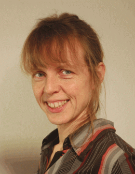
Prof. Dr. Friederike Schmid
Johannes Gutenberg University Mainz - Institute of PhysicsResearch Mission
Our research is devoted to the statistical physics of soft matter, with special interest in polymers and membranes. Within the IMPRS, we are interested in learning how such concepts can be help to better understand biological materials and cellular processes.
Experimental Techniques
We combine numerical calculations, computer simulations, and analytical theory. Much effort is also spent on the development of new efficient simulation techniques and multiscale simulation methods.

Prof. Dr. Erin Schuman
MPI for Brain Research - Synaptic PlasticityNeurobiology
Research Mission
Erin Schuman lab’s long-standing research interest is the study of cellular mechanisms and neural circuits that underlie information processing and storage. The lab focuses on the molecular and cell biological processes that control protein synthesis and degradation in neurons and their synapses.
Experimental Techniques
mRNA sequencing, ribosome profiling, proteomics, in situ hybridization, super-resolution imaging, bioinformatics, electrophysiology, cryo-electron tomography, biochemistry, molecular biology, fluorescence-activated synaptosome sorting, BONCAT, FUNCAT, polysome profiling, proximity-labelling cultured rodent neurons, brain slices, heterologous cells
Selected Publications
The proteomic landscape of synaptic diversity across brain regions and cell types.
Cell 2023
An abundance of free regulatory (19 S) proteasome particles regulates neuronal synapses.
Science 2023
Neuronal ribosomes exhibit dynamic and context-dependent exchange of ribosomal proteins.
Nature Communications 2021
Monosomes actively translate synaptic mRNAs in neuronal processes.
Science 2020
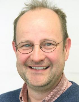
Prof. Dr. Harald Schwalbe
Goethe University Frankfurt - Institute for Organic Chemistry and Chemical BiologyResearch Mission
My group conducts research in integrated structural biology to study structure and dynamics of proteins, RNAs, and DNAs and their interaction. We are interested in understanding conformational dynamics of RNA structures and collaborate with MD scientists. Since 2020, a major focus is on SARS-CoV-2, including new inhibitions and drugs. We are interested in time-resolved NMR spectroscopy, especially light-induced methods
Experimental Techniques
Our main focus is on NMR spectroscopy. In addition, we solve X-ray structures and single particle cryo-EM.
Selected Publications
Three-state mechanism couples ligand and temperature sensing in riboswitches
Nature 2013
Secondary structure determination of conserved SARS-CoV-2 RNA elements by NMR spectroscopy
Nucl. Acids Res. 2000
Exploring the Druggability of Conserved RNA Regulatory Elements in the SARS-CoV-2 Genome
Angew. Chem. 2021
Cysteine oxidation and disulfide formation in the ribosomal exit tunnel
Nat. Comm. 2020
Synonymous Codons Direct Cotranslational Folding toward Different Protein Conformations
Mol. Cell 2016

Dr. Lukas Stelzl
Johannes Gutenberg University Mainz - Institute of Molecular Biology (IMB)Biophysics
Research Mission
Our aim is to understand how liquid-liquid phase separation and biomolecular condensates provide for specific regulation in the cell and how this regulation is lost in aging and disease. We are elucidating how molecular recognition of disordered biomolecules gives rise to specific regulation in (post)-transcriptional regulation of genes. We are studying how phase separation can be a robust mechanism and how it becomes dysregulated in diseases.
Experimental Techniques
To understand disordered biomolecules, their assemblies, condensates and phase behavior we are employing multi-scale simulations. With coarse-grained simulations, we study self-assembly and phase behavior and use all-atom simulations to resolve molecular interactions with atomic resolution. We are developing multi-scale methods further to tackle challenging biological problems. With Bayesian approaches we combine simulation and experiments.
Selected Publications
Global Structure of the Intrinsically Disordered Protein Tau Emerges from Its Local Structure
JACS Au 2022
Disease-linked TDP-43 hyperphosphorylation suppresses TDP-43 condensation and aggregation
EMBO J 2022
Local frustration determines loop opening during the catalytic cycle of an oxidoreductase
eLife 2020
Kinetics from Replica Exchange Molecular Dynamics Simulations
J Chem Theory Comput 2017
Resolving the Conformational Dynamics of DNA with Ångstrom Resolution by Pulsed Electron–Electron Double Resonance and Molecular Dynamics
J Am Chem Soc 2017

Dr. Till Stephan
Goethe University Frankfurt - Institute of BiochemistryCell Biology
Research Mission
Cells rely on specialized organelles and subcellular structures, each with a distinct architecture finely tuned to its specific function. Our research employs advanced imaging techniques to investigate the mechanisms that shape the architecture of eukaryotic organelles at the nanometer scale. In particular, we focus on mitochondria and the endoplasmic reticulum and their function in energy conversion or metabolism.
Experimental Techniques
We employ advanced light microscopy, including STED super-resolution microscopy, to study the nanoscale architecture of eukaryotic (preferentially human) cells. For fluorescence labeling, we utilize approaches like CRISPR/Cas9 genome editing. Our work is typically embedded in interdisciplinary collaborations that combine fluorescence imaging with electron microscopy and biochemical analyses to uncover organelle structure and function.
Selected Publications
MICOS assembly controls mitochondrial inner membrane remodeling and crista junction redistribution to mediate cristae formation
The EMBO Journal (EMBOJ) 2020
Multicolor 3D MINFLUX nanoscopy of mitochondrial MICOS proteins
Proceedings of the National Academy of Sciences (PNAS) 2020
Multi-color live-cell STED nanoscopy of mitochondria with a gentle inner membrane stain
Proceedings of the National Academy of Sciences (PNAS) 2022
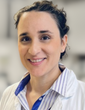
Dr. Alexandra Stolz
Goethe University Frankfurt - Institute of Biochemistry II (IBC2)Autophagy
Research Mission
Cellular homeostasis is preserved by maintenance and stress response pathways. Upon them is autophagy, a degradation pathway with disease relevance and responsible for the turnover of individual proteins as well as whole organelles like the endoplasmic reticulum (ER). We are interested in the regulation of and molecular mechanisms within selective autophagy pathways of the ER and aim to develop chemical modulators for disease treatment.
Experimental Techniques
As cellular models, we mostly use human and mouse cell lines. Our diverse methodological repertoire includes biochemical and cell biological methods such as cell culture, a variety of standard biochemical assays, immuno-precepetation, live-cell imaging, confocal imaging and mass spectrometry.
Selected Publications
The function of ER-phagy receptors is regulated through phosphorylation-dependent ubiquitination pathways.
Nat Commun. 2023
Role of FAM134 paralogues in endoplasmic reticulum remodeling, ER-phagy, and Collagen quality control.
EMBO Rep. 2021
An mTOR-independent Macroautophagy Activator Ameliorates Tauopathy and Prionopathy Neurodegeneration Phenotypes
bioRxiv 2024

Dr. Beata Turoňová
MPI of Biophysics - Molecular SociologyStructural Biology
Research Mission
We aim to facilitate in situ structural analysis by developing novel algorithms for cryo electron tomography and subtomogram averaging. Our method development focuses on improving tomogram quality and integrating contextual information into subtomogram averaging routines. The goal is to allow for reliable and objective analysis of tomograms as well as macromolecular complexes.
Experimental Techniques
The main focus is software method development. We use cryo electron tomography, often in combination with focused ion-beam milling, to obtain data for the method development. To ensure their robustness, the methods are tested on a variety of samples from virus-like particles (e.g. HIV, Sars-Cov-2) and real viruses (e.g. Sars-Cov-2, vaccinia) to very large macromolecular complexes (e.g. nuclear pore complexes).
Selected Publications
In situ structural analysis of SARS-CoV-2 spike reveals flexibility mediated by three hinges
Science 2020
Benchmarking tomographic acquisition schemes for high-resolution structural biology
Nature Communications 2020
Cone-shaped HIV-1 capsids are transported through intact nuclear pores
Cell 2021

Dr. Janet Vonck
MPI of Biophysics - Structural BiologyStructural Biology
Research Mission
Membrane proteins play essential roles in many cellular processes including transport, creating and maintaining ion gradients, and signaling, between cellular compartments and the cell and its environment. But our knowledge of their structure is still far behind that of soluble proteins. We study the structure and dynamics of membrane proteins and membrane protein complexes by cryo-EM.
Experimental Techniques
We use the pipeline of single-particle cryo-EM from sample vitrification, microscope data collection, image processing and particle reconstruction to model building.
Selected Publications
High-resolution structure and dynamics of mitochondrial complex I – insights into the proton pumping mechanism
Science Advances 7, eabj3221 2021
Structural basis of proton-coupled potassium transport in the KUP family
Nature Communications 11, 626 2020
Structure, mechanism, and regulation of the chloroplast ATP synthase
Science 360, eaat4318 2018

Dr. Sonja Welsch
MPI of BiophysicsResearch Mission
A thorough analysis of structural elements within cells is key to understanding the function of cellular machines. Electron microscopy (EM) and tomography (ET) are pivotal in generating high-resolution information of cellular ultrastructure and macromolecular organization. We support researchers and projects by maintaining and providing access to state-of-the-art EM equipment and by providing training that is tailored to each researcher’s needs.
Experimental Techniques
We use EM and ET as well as correlative fluorescence and electron microscopy techniques to study biological materials fixed by vitrification in a close-to-native state and imaged under cryo-conditions.

Dr. Florian Wilfling
MPI of BiophysicsCell Biology
Research Mission
Proteins are workhorses of cells and perform a vast array of functions. Often this requires the formation of large multi-protein complexes which act as a macromolecular machine for individual tasks. How disused, damaged or misassembled macromolecular machines are disposed is largely unknown. Our main goal is to systematically identify signals and factors important for surveillance of the assembly and functional state of macromolecular machines.
Experimental Techniques
We integrate a variety of techniques including biochemistry, advanced live cell microscopy, state-of-the-art quantitative mass spectrometry, and unbiased system-wide approaches to study the cell biology of selective autophagy. A particular focus of our group is to use a correlative cryo-electron tomography approach to zoom into the structural details of autophagy in cells to gain a molecular understanding of this process in health and disease.
Selected Publications
Selective autophagy degrades nuclear pore complexes
Nature Cell Biology 2020
A selective autophagy pathway for phase separated endocytic protein deposits
Molecular Cell 2020
In situ structural analysis reveals membrane shape transitions during autophagosome formation.
Proceedings of the National Academy of Sciences of the United States of America 2022
Autophagy preferentially degrades non-fibrillar polyQ aggregates.
Molecular Cell 2024
Selective autophagy of macromolecular complexes: what does it take to be taken?
Journal of Molecular Biology 2024

Dr. Martin Beck
MPI of Biophysics - Molecular SociologyStructural Biology
Research Mission
Functional cellular modules have often been characterised in vitro. However, relatively little is known about their interplay and spatio-temporal arrangement in the context of living cells, i.e. their 'molecular sociology'. We use integrative, in situ structural biology techniques to study the structure, function and assembly of very large macromolecular complexes in their native environment.
Experimental Techniques
We rely on a diverse methodological repertoire, including cryo-EM, structural proteomics, biochemistry, imaging, animal model systems and computational modeling

Prof. Dr. Ivan Dikic
Goethe University Frankfurt - Institute of Biochemistry II (IBC2)Biochemistry
Research Mission
Cellular homeostasis is achieved, among others, by tight regulation of the two major degradation pathways – the ubiquitin-proteasome system (UPS) and autophagy. It is critical to understand the structure and function of individual components within each of these pathways, as any disruption to either of the processes can lead to neurodegenerative diseases, cancer and other pathologies. To study functional and structural properties of components of
Experimental Techniques
To tackle our research questions, we rely on a wide variety of biological and chemical techniques, including biochemistry, proteomics, cell imaging, X-ray crystallography, Cryo-EM and animal model systems.

Prof. Dr. Dorothee Dormann
Johannes Gutenberg University Mainz - Institute of Molecular Biology (IMB)Neurodegeneration
Research Mission
Our research focuses on the molecular mechanisms of RNA-binding protein (RBP) dysfunction in neurodegenerative diseases, most notably the currently incurable disorders ALS (amyotrophic lateral sclerosis), FTD (frontotemporal dementia), and Alzheimer's disease. We aim to understand how specific, disease-linked RBPs, such as TDP-43 and FUS, mislocalize and aggregate in these disorders, to provide a mechanistic basis for new therapeutic strategies.
Experimental Techniques
We dissect the complex pathomechanism using a reductionist approach based on in vitro reconstitution and cellular experiments. We use recombinant proteins, protein/RNA biochemistry and biophysics to understand molecular events. As cellular models, we mostly use mammalian cell lines. We use techniques such as confocal microscopy, live cell imaging, protein-RNA interaction and phase separation assays, and employ genomics and proteomics approaches.
Selected Publications
Disease-linked TDP-43 hyperphosphorylation suppresses TDP-43 condensation and aggregation
EMBO Journal 2022
Phase Separation of FUS Is Suppressed by Its Nuclear Import Receptor and Arginine Methylation
Cell 2018
Adding intrinsically disordered proteins to biological ageing clocks
Nature Cell Biology 2024

Prof. Dr. Achilleas Frangakis
Goethe University Frankfurt - Institute of BiophysicsBioimaging
Research Mission
Cell adhesion is an essential process that allows for cells to attach and interact with other cells and their environment in order to perform a multitude of tasks. Although adhesion is directly related to infection processes and disease, it is currently only poorly understood. Our mission is to study and decipher the adhesion mechanisms of both eukaryotic and bacterial cells.
Experimental Techniques
We use a large arsenal of different technologies to complement our primary methodologies of cryo-electron tomography, image processing and machine learning. Our main model system to study bacterial adhesion is M. pneumoniae & genitalium. For eukaryotic systems we utilise various endothelial and epithelial cell lines.

Prof. Dr. Ana García Sáez
MPI of BiophysicsBioimaging
Research Mission
Our department aims at understanding the underlying physical principles and molecular mechanisms that govern membrane organization and dynamics. A key open question is how membrane behavior and signaling are coordinated by the interplay between molecular components and the membrane properties. We focus our research on the mitochondrial alterations during apoptosis as well as on membrane permeabilization as a common theme in regulated cell death.
Experimental Techniques
We have established a multi-disciplinary department that develops new microscopy tools and combines biophysics, biochemistry and cell biology in reconstituted minimal systems and in single cells to analyze mitochondrial permeabilization and additional alterations during apoptosis from a quantitative perspective. We also apply these methods to study the molecular mechanisms of membrane permeabilization that execute other forms of cell death.
Selected Publications
The interplay between BAX and BAK tunes apoptotic pore growth to control mitochondrial DNA-mediated inflammation
Molecular Cell 2022
DRP1 interacts directly with BAX to induce its activation and apoptosis
EMBO Journal 2022
BCL-2-family protein tBID can act as a BAX-like effector of apoptosis
EMBO Journal 2022

Dr. Bernhard Hampölz
MPI of Biophysics - Molecular SociologyCell Biology
Research Mission
Our aim is to understand how macromolecular complexes and in particular the Nuclear Pore Complex (NPC) assemble in the context of animal development. To that end we use embryos and the female germline of the fruitfly Drosophila melanogaster to delineate principles of how NPCs form in a multicellular organism and how their assembly relies on the major regulatory inputs of early development.
Experimental Techniques
Our methodological repertoire is broad and spans techniques as diverse as cultivating and crossing fruitfly lines (aka “Fly pushing”) and in vitro biochemistry. This spectrum contains Drosophila genetics, general cell biological techniques, organ dissection, plastic and Cryo-EM (including CLEM). Our core technology is imaging of living Drosophila embryos, ovaries and larval brains.
Selected Publications
Nuclear Pores Assemble from Nucloporin Condensates During Oogenesis
Cell 2019
Pre-assembled Nuclear Pores Insert Into the Nuclear Envelope During Early Development
Cell 2016
The small GTPase Ran defines Nuclear Pore Complex Asymmetry
Cell 2025

Prof. Dr. Inga Hänelt
Goethe University Frankfurt - Institute of BiochemistryBiochemistry
Research Mission
Proteins involved in substrate translocation across biological membranes are diverse and the variety that nature has evolved to control substrate flux absolutely fascinates us. A major focus of my group is on bacterial membrane transport systems and, in particular, potassium transport systems that are essential for the survival of a single cell and a bacterial community, namely biofilms.
Experimental Techniques
Our lab applies a variety of methods, including biochemistry, structural biology, EPR spectroscopy, microbiology and electrophysiology for understanding the molecular principles of membrane transport proteins.

Prof. Dr. Mike Heilemann
Goethe University Frankfurt - Institute of Physical and Theoretical ChemistryBioimaging
Research Mission
Our mission is to develop super-resolution fluorescence microscopy into an "optical omics" technology for structural cell biology. We develop imaging tools for quantitative microscopy with single-protein resolution, along with novel computational tools for microscopy. We investigate how proteins organize in cells and tissue, and how this organization correlates with protein function.
Experimental Techniques
super-resolution microscopy (dSTORM, PALM, DNA-PAINT, STED, SOFI), live cell microscopy, deep-learning assisted microscopy, chemical biology, fluorescence spectroscopy

Prof. Dr. Gerhard Hummer
MPI of Biophysics - Theoretical BiophysicsBiophysics
Research Mission
We use molecular dynamics simulations and integrative modeling to study biological systems from molecular to organellar scales. Focus areas include the dynamics of the nuclear pore complex; membrane remodeling in cell homeostasis, autophagy and viral infection; and membrane channel and transporter function. To tackle issues of size and complexity in molecular dynamics simulations at organellar scales, we develop novel simulation and AI methods.
Experimental Techniques
We use a wide range of computational and theoretical techniques including molecular dynamics simulations; integrative modeling; artificial intelligence / machine learning; statistical physics; computational physics; theoretical chemistry; quantum chemistry; computational chemistry; bioinformatics; high-performance computation; kinetic modeling; stochastic processes; and Bayesian statistics.
Selected Publications
Artificial intelligence reveals nuclear pore complexity
bioRxiv 2022
Global structure of the intrinsically disordered protein tau emerges from its local structure
JACS Au 2022
Membrane fusion and drug delivery with carbon nanotube porins
Proc. Natl. Acad. Sci. U.S.A. 2021
FAM134B-RHD protein clustering drives spontaneous budding of asymmetric membranes
J. Phys. Chem. Lett. 2022
In situ structural analysis of SARS-CoV-2 spike reveals flexibility mediated by three hinges
Science 2021

Dr. Eugene Kim
MPI of BiophysicsBiophysics
Research Mission
Cellular DNA is highly organized not only to fit within the cells, but also to regulate genomic processes. Yet, what determines DNA organization and how it impacts on cellular function remain largely unknown. We study biochemical and physical processes underlying chromosome organization by monitoring their dynamics at the single-molecule level with high spatiotemporal resolution.
Experimental Techniques
We employ single-molecule fluorescence imaging, single-molecule force spectroscopy, and correlative light and electron microscopy techniques, in combination with analytical biochemistry tools.

Dr. Melanie McDowell
MPI of BiophysicsStructural Biology
Research Mission
Almost all eukaryotic membrane proteins start their life in the cytosol and must journey to the cellular membrane where they function. Multiple pathways are employed to recognise different classes of membrane proteins, deliver them to the correct membrane and insert them into the lipid bilayer. We strive to understand the protein factors involved in these pathways at the endoplasmic reticulum, the primary destination for most membrane proteins.
Experimental Techniques
We use an integrative approach to study protein structure, interactions and function. Techniques include cryo-EM, x-ray crystallography, biophysical methods, in vitro biochemical reconstitution and pull-outs of native protein complexes from model organisms such as yeast and Chaetomium thermophilum.
Selected Publications
Structural and molecular mechanisms for membrane protein biogenesis by the Oxa1 superfamily
Nature Structural and Molecular Biology 2021
Structural basis of tail-anchored membrane protein biogenesis by the GET insertase complex
Molecular Cell 2020

Prof. Dr. Christian Münch
Goethe University Frankfurt - Institute of Biochemistry II (IBC2)Cell Biology
Research Mission
Cells respond to stress with highly coordinated and dynamic responses. Their full complexity and spatio-temporal control remains poorly understood. We focus on a range of stresses - (mitochondrial) protein misfolding, infection, cancer - to study their effects on a systems biology/medicine level. We aim to define the molecular details of stress responses, their roles in diseases, and potential avenues for therapeutic intervention.
Experimental Techniques
We employ biochemical, cell biological, genetic, and proteomics approaches to comprehensively monitor the multiple facets of stress responses. To grasp dynamic, cell-wide changes, we develop proteomics methods, such as for translation & degradation. We then apply computational approaches on our multi-level data to gain integrated systems information. Our work largely focuses on mammalian cell culture systems and clinical models.
Selected Publications
A cytosolic surveillance mechanism activates the mitochondrial UPR
Nature 2023
Global mitochondrial protein import proteomics reveal distinct regulation by translation and translocation machinery
Molecular Cell 2022
Protein import motor complex reacts to mitochondrial misfolding by reducing protein import and activating mitophagy
Nature Commun 2022
Proteomics of SARS-CoV-2-infected host cells reveals therapy targets
Nature 2020
Functional Translatome Proteomics Reveal Converging and Dose-Dependent Regulation by mTORC1 and eIF2α
Molecular Cell 2020

Dr. Bonnie Murphy
MPI of BiophysicsStructural Biology
Research Mission
Our group is focused on gaining a better understanding of the structure and especially the mechanism of redox and metalloproteins across the tree of life. We also work to develop methods for elemental mapping in macromolecular complexes, combining analytical techniques typically used for dose-tolerant inorganic samples with the image processing tools of single-particle analysis.
Experimental Techniques
We use single-particle cryo-EM as a primary tool in the lab, complemented by biochemical and electrochemical tools. We use additional analytical capabilities of the microscope in STEM mode (eg. 4D-STEM, EELS analysis), giving us richer information about our sample.
Selected Publications
see our website / Google scholar page for up-to-date publication list
2026

Dr. Beata Turoňová
MPI of Biophysics - Molecular SociologyStructural Biology
Research Mission
We aim to facilitate in situ structural analysis by developing novel algorithms for cryo electron tomography and subtomogram averaging. Our method development focuses on improving tomogram quality and integrating contextual information into subtomogram averaging routines. The goal is to allow for reliable and objective analysis of tomograms as well as macromolecular complexes.
Experimental Techniques
The main focus is software method development. We use cryo electron tomography, often in combination with focused ion-beam milling, to obtain data for the method development. To ensure their robustness, the methods are tested on a variety of samples from virus-like particles (e.g. HIV, Sars-Cov-2) and real viruses (e.g. Sars-Cov-2, vaccinia) to very large macromolecular complexes (e.g. nuclear pore complexes).
Selected Publications
In situ structural analysis of SARS-CoV-2 spike reveals flexibility mediated by three hinges
Science 2020
Benchmarking tomographic acquisition schemes for high-resolution structural biology
Nature Communications 2020
Cone-shaped HIV-1 capsids are transported through intact nuclear pores
Cell 2021